Rib Cage Muscles Labeled / Thorax Wikipedia : 2) innervated by the 12 pairs of thoracic spinal nerves.. There are five muscles that make up the thoracic cage; Our latest youtube film is ready to run. The best way to learn anatomy is to repeat as much as you can. The first seven ribs in the rib cage are attached to the sternum by pliable cartilages called costal cartilages; The rectus abdominis is an important postural muscle.
Scalene and latissimus dorsi muscles in the upper back also assist with raising the shoulder blade to add extra space in the rib cage. The heads of ribs 1, 10, 11, and 12 have a single facet for articulation with the bodies of the thoracic vertebrae. With the upper ribs, closer to the nodule (and in the case of lower ribs, a little further from the nodule) they are curved and have a rough surface that connects them with muscles, angulus costae. Check spelling or type a new query. Rib cage muscles labeled :
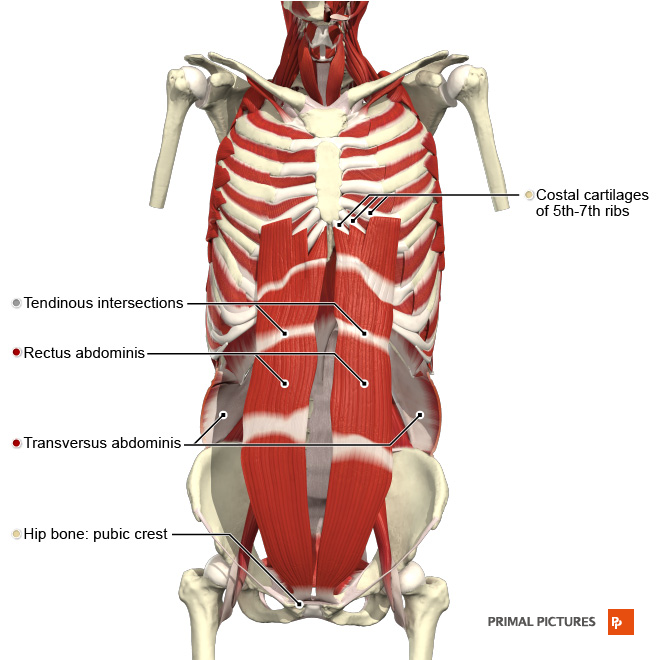
1 from There are two parallel muscles, separated by a midline band of connective tissue called the linea alba. In comparison, the first two ribs are shorter and more curved. Check spelling or type a new query. Below the 2nd rib, laterally. It furthermore supports breathing and stabilizes the. Knowing what can affect your rib cage, back muscles, and ligaments that support the spine can help to take steps to relieve the pain. It provides a strong framework onto which the muscles of the shoulder girdle, chest, upper abdomen and back can attach. Each are symmetrically paired on a right and left side.
Of the remaining five ribs, which are called false, the first three have their costal cartilages connected to the cartilage above them.
Quizzes are the secret to your success! The rib cage has three important functions: The rib cage is the arrangement of ribs attached to the vertebral column and sternum in the thorax of most vertebrates that encloses and protects the vital organs such as the heart, lungs and great vessels. It is also the center around which the superior 10 ribs directly or indirectly attached. Skull, vertebral column, rib cage appendicular: Serratus posterior and pectoralis minor muscles assist with raising the upper ribs. Intercostal muscles function area course human anatomy kenhub youtube / check spelling or type a new query. 2) innervated by the 12 pairs of thoracic spinal nerves. The most common causes of rib cage pain are a pulled muscle or bruised ribs. The best way to learn anatomy is to repeat as much as you can. The pectoralis major muscles (also known as the pecs) are located on the front of the rib cage. The human rib cage is made up of 12 paired rib bones; Knowing what can affect your rib cage, back muscles, and ligaments that support the spine can help to take steps to relieve the pain.
With the upper ribs, closer to the nodule (and in the case of lower ribs, a little further from the nodule) they are curved and have a rough surface that connects them with muscles, angulus costae. Anatomy of the chest muscles. Check spelling or type a new query. Anatomy of the rib cage diagram. These muscles act to change the volume of the thoracic cavity during respiration.
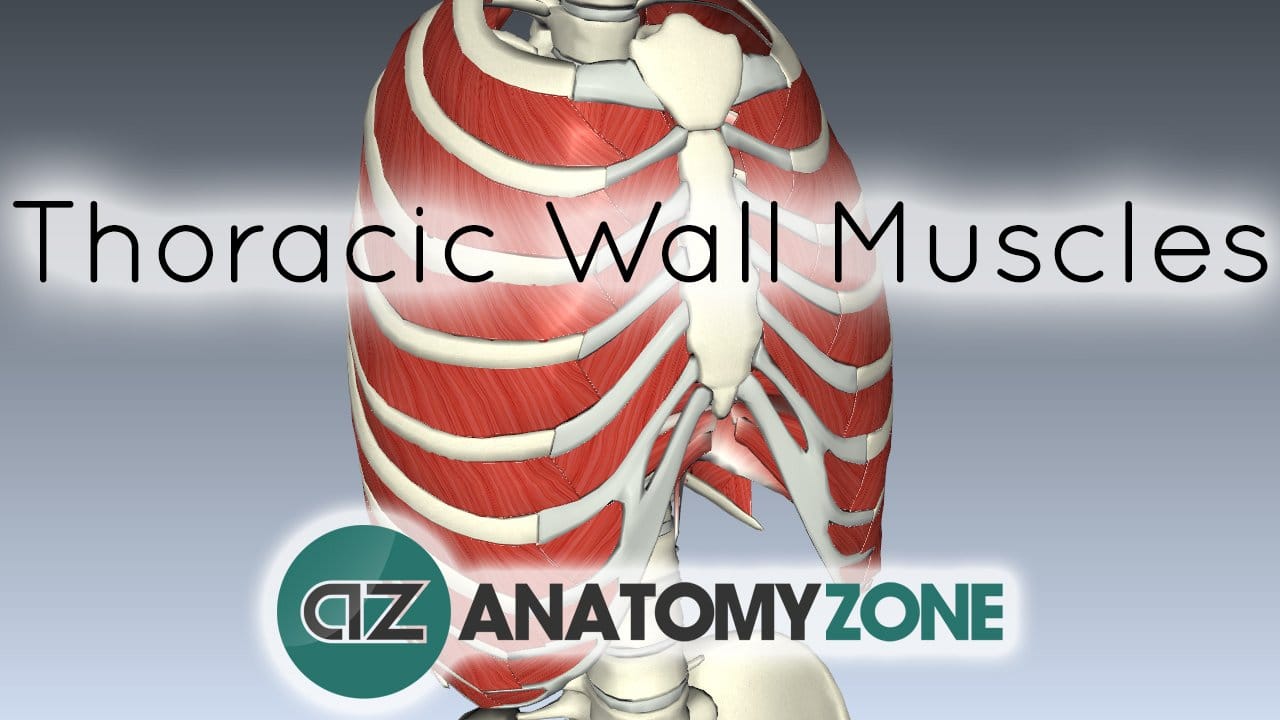
Muscles Of The Thoracic Wall 3d Models Video Tutorials Notes Anatomyzone from anatomyzone.com The average skeleton contains 24 individual ribs, formed in 12. There are two parallel muscles, separated by a midline band of connective tissue called the linea alba. In this image, you will find thoracic vertebrum, costochondral joint, costal cartilage, costal margin, costal arch, thoracic vertebrum, xiphoid process, xiphisternal joint, body, manubrial sternal joint, manubrium, the sternal notch in it. Below the 2nd rib, laterally. There are five muscles that make up the thoracic cage; The intercostals (external, internal and innermost), subcostals, and transversus thoracis. Ribs 11 and 12 do not have necks or tubercles and the anterior tips of. The rib cage consists of 24 ribs, 12 on either side, and it shields the organs of the chest, including the heart and the lungs, from damage.
11 muscles in between each rib
These muscles act to change the volume of the thoracic cavity during respiration. The body, or shaft, of the rib is thin, flat and curved. The costal angle also marks the attachment for some of the deep back muscles to. The pecs attach to the humerus near the shoulder joint and originate on the breastbone in the center of the chest. The most common causes of rib cage pain are a pulled muscle or bruised ribs. The curve becomes most prominent at the costal angle, which is when the rib turns anterolaterally. Rib cage muscles labeled : The first seven ribs in the rib cage are attached to the sternum by pliable cartilages called costal cartilages; Intercostal muscles function area course human anatomy kenhub youtube / check spelling or type a new query. Rib 1 is also flattened horizontally. Skull, vertebral column, rib cage appendicular: Maybe you would like to learn more about one of these? Scalene and latissimus dorsi muscles in the upper back also assist with raising the shoulder blade to add extra space in the rib cage.
The heads of ribs 1, 10, 11, and 12 have a single facet for articulation with the bodies of the thoracic vertebrae. The best way to learn anatomy is to repeat as much as you can. Aalso known as the six pack, is a paired muscle running vertically on each side of the front wall of the abdomen. The sternocleidomastoid muscle, which comes from the jaw and crosses over the neck, moves the breastbone upward. The rib cage consists of 24 ribs, 12 on either side, and it shields the organs of the chest, including the heart and the lungs, from damage.
Muscles Of Respiration Physiopedia from www.physio-pedia.com The ribs are attached to the breastbone, which is the. Skull, vertebral column, rib cage appendicular: Intercostal muscles function area course human anatomy kenhub youtube / check spelling or type a new query. Our latest youtube film is ready to run. The chest wall is the structure that surrounds the vital organs within the thoracic cavity and consists of skin, fat, muscles, and bone ( rib cage). Each are symmetrically paired on a right and left side. 2) innervated by the 12 pairs of thoracic spinal nerves. And upper arms during movement.
The rib cage consists of 24 ribs, 12 on either side, and it shields the organs of the chest, including the heart and the lungs, from damage.
The rib cage has three important functions: Of the remaining five ribs, which are called false, the first three have their costal cartilages connected to the cartilage above them. 16 photos of the rib cage diagram with organs. With the upper ribs, closer to the nodule (and in the case of lower ribs, a little further from the nodule) they are curved and have a rough surface that connects them with muscles, angulus costae. Raises rib cage during inhalation. 1)elevate the rib cage, increasing its anterior dimension and lung volume. Anatomy of the chest muscles. Knowing what can affect your rib cage, back muscles, and ligaments that support the spine can help to take steps to relieve the pain. Other causes of pain in the rib cage area may include: Scalene and latissimus dorsi muscles in the upper back also assist with raising the shoulder blade to add extra space in the rib cage. The upper edge is round and the lower sharp. The pecs attach to the humerus near the shoulder joint and originate on the breastbone in the center of the chest. 2) innervated by the 12 pairs of thoracic spinal nerves.
It is responsible for pulling the rib cage toward the pelvis rib cage muscles. 11 muscles in between each rib


0 Komentar